
HL Paper 3
The linear attenuation coefficient μ of a material is affected by the energy of the X-ray beam and by the density ρ of the material. The mass absorption coefficient is equal to to take into account the density of the material.
The graph shows the variation of mass absorption coefficient with energy of the X-ray beam for both muscle and bone.

Show that the attenuation coefficient for bone of density 1800 kg m–3, for X-rays of 20 keV, is about 7 cm–1.
The density of muscle is 1200 kg m–3. Calculate the ratio of intensities to compare, for a beam of 20 keV, the attenuation produced by 1 cm of bone and 1 cm of muscle.
Suggest why more energetic beams of about 150 keV would be unsuitable for imaging a bone–muscle section of a body.
The density of muscle is 1075 kg m–3 and the speed of ultrasound in muscle is 1590 m s–1.
State a typical frequency used in medical ultrasound imaging.
Describe how an ultrasound transducer produces ultrasound.
Calculate the acoustic impedance Z of muscle.
Ultrasound of intensity 0.012 Wcm–2 is incident on a water–muscle boundary. The acoustic impedance of water is 1.50 x 10⁶ kgm–2s–1.
The fraction of the incident intensity that is reflected is given by
where Z1 and Z2 are the acoustic impedances of medium 1 and medium 2.
Calculate the intensity of the reflected signal.
In the context of nuclear magnetic resonance (NMR) imaging explain the role of
Outline why the fracture in a broken bone can be seen in a medical X-ray image.
The diagram shows X-rays incident on tissue and bone.

The thicknesses of bone and tissue are both 0.054 m.
The intensity of X-rays transmitted through bone is Ib and the intensity transmitted through tissue is It.
The following data are available.
Mass absorption coefficient for bone = mass absorption
coefficient for tissue = 1.2 × 10⁻²m2kg–1
Density of bone = 1.9 × 10³ kgm–3
Density of tissue = 1.1 × 10³ kgm–3
Calculate the ratio .
the large uniform magnetic field applied to the patient.
the radio-frequency signal emitted towards the patient.
the non-uniform magnetic field applied to the patient.
The table shows the speed of ultrasound and the acoustic impedance for different media.
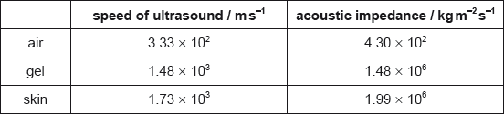
The fraction F of the intensity of an ultrasound wave reflected at the boundary between two media having acoustic impedances Z1 and Z2 is given by F = .
Outline how ultrasound is generated for medical imaging.
Describe one advantage and one disadvantage of using high frequencies ultrasound over low frequencies ultra sound for medical imaging.

Suggest one reason why doctors use ultrasound rather than X-rays to monitor the development of a fetus.
Calculate the density of skin.
Explain, with appropriate calculations, why a gel is used between the transducer and the skin.
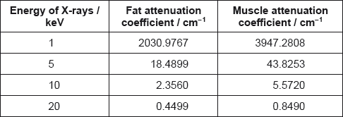
The attenuation values for fat and muscle at different X-ray energies are shown.
Outline the formation of a B scan in medical ultrasound imaging.
State what is meant by half-value thickness in X-ray imaging.
A monochromatic X-ray beam of energy 20 keV and intensity I0 penetrates 5.00 cm of fat and then 4.00 cm of muscle.
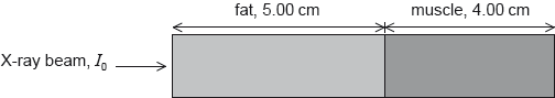
Calculate, in terms of I0, the final beam intensity that emerges from the muscle.
Compare the use of high and low energy X-rays for medical imaging.
Some optic fibres consist of a core surrounded by cladding as shown in the diagram.

Calculate the maximum angle β for light to travel through the fibre.
Refractive index of core = 1.50
Refractive index of cladding = 1.48
Outline how the combination of core and cladding reduces the overall dispersion in the optic fibres.
The photograph shows an X-ray image of a hand.
 © International Baccalaureate Organization 2020.
Explain how attenuation causes the contrast between soft tissue and bone in the image.
X-ray images of other parts of the body require the contrast to be enhanced. State one technique used in X-ray medical imaging to enhance contrast.
In nuclear magnetic resonance imaging (NMR) a patient is exposed to a strong external magnetic field so that the spin of the protons in the body align parallel or antiparallel to the magnetic field. A pulse of a radio frequency (RF) electromagnetic wave is then directed at the patient.
Describe the effect of the RF signal on the protons in the body.
Outline the measurement that needs to be made after the RF signal is turned off.
Describe how the measurement in (b) provides diagnostic information for the doctor.
Outline how ultrasound, in a medical context, is produced.
Suggest the advantage in medical diagnosis of using ultrasound of frequency 1 MHz rather than 0.1 MHz.
Ultrasound can be used to measure the dimensions of a blood vessel. Suggest why a B scan is preferable to an A scan for this application.
An X-ray beam of intensity I0 is incident on lead. After travelling a distance x through the lead the intensity of the beam is reduced to I.
The graph shows the variation of ln with x.

Show that the attenuation coefficient of lead is 60 cm–1.
A technician operates an X-ray machine that takes 100 images each day. Estimate the width of the lead screen that is required so that the total exposure of the technician in 250 working days is equal to the exposure that the technician would receive from one X-ray exposure without the lead screen.
An ultrasound A-scan is performed on a patient.
The graph shows a received signal incident upon a transducer to produce an A-scan. The density of the soft tissue being examined is approximately 1090 kg m-3.

State one advantage and one disadvantage of using ultrasound imaging in medicine compared to using x-ray imaging.
Advantage:
Disadvantage:
Suggest why ultrasound gel is necessary during an ultrasound examination.
Ultrasound of intensity 50 mW m-2 is incident on a muscle. The reflected intensity is 10 mW m-2. Calculate the relative intensity level between the reflected and transmitted signals.
The acoustic impedance of soft tissue is 1.65 × 10⁶ kg m-2 s-1. Show that the speed of sound in the soft tissue is approximately 1500 m s–1.
Estimate, using data from the graph, the depth of the organ represented by the dashed line.
In the ultrasound scan the frequency is chosen so that the distance between the transducer and the organ is at least 200 ultrasound wavelengths. Estimate, based on your response to (b)(ii), the minimum ultrasound frequency that is used.
A physician has a range of frequencies available for ultrasound. Comment on the use of higher frequency sound waves in an ultrasound imaging study.
A beam of ultrasound of intensity I0 enters a layer of muscle of thickness 4.1 cm.

The fraction of the intensity that is reflected at a boundary is
where Z1 and Z2 are the acoustic impedances of the two media at the boundary. After travelling a distance x in a medium the intensity of ultrasound is reduced by a factor e–μx where μ is the absorption coefficient.
The following data are available.
Acoustic impedance of muscle = 1.7 × 10⁶ kg m–2 s–1
Acoustic impedance of bone = 6.3 × 10⁶ kg m–2 s–1
Absorption coefficient of muscle = 23 m–1
Determine, in terms of I0, the intensity of ultrasound that is incident on the muscle–bone boundary.
Determine, in terms of I0, the intensity of ultrasound that is reflected at the muscle–bone boundary.
Determine, in terms of I0, the intensity of ultrasound that returns to the muscle–gel boundary.
Explain the cause of the radio-frequency emissions from a patient’s body during nuclear magnetic resonance (NMR) imaging.
Outline how a gradient field allows NMR to be used in medical resonance imaging.
Identify one advantage of NMR over ultrasound in medical situations.
A parallel beam of X-rays travels through 7.8 cm of tissue to reach the bowel surface. Calculate the fraction of the original intensity of the X-rays that reach the bowel surface. The linear attenuation coefficient for tissue is 0.24 cm–1.
The fluid in the bowel has a similar linear attenuation coefficient as the bowel surface. Gases have much lower linear attenuation coefficients than fluids. Explain why doctors will fill the bowel with air before taking an X-ray image.
An X-ray beam, of intensity , is used to examine the flow of blood through an artery in the leg of a patient. The beam passes through an equal thickness of blood and soft tissue.

The thickness of blood and tissue is 5.00 mm. The intensity of the X-rays emerging from the tissue is and the intensity emerging from the blood is .
The following data are available.
Mass absorption coefficient of tissue = 0.379 cm2 g–1
Mass absorption coefficient of blood = 0.385 cm2 g–1
Density of tissue = 1.10 × 10³ kg m–3
Density of blood = 1.06 × 10³ kg m–3
Show that the ratio is close to 1.
State and explain, with reference to you answer in (a)(i), what needs to be done to produce a clear image of the leg artery using X-rays.
In nuclear magnetic resonance (NMR) protons inside a patient are made to emit a radio frequency electromagnetic radiation. Outline the mechanism by which this radiation is emitted by the protons.
State the property of protons used in nuclear magnetic resonance (NMR) imaging.
Explain how a gradient field and resonance are produced in NMR to allow for the formation of images at a specific plane.
The diagram represents a simple optical astronomical reflecting telescope with the path of some light rays shown.

It is proposed to build an array of radio telescopes such that the maximum distance between them is 3800 km. The array will operate at a wavelength of 2.1 cm.
Comment on whether it is possible to build an optical telescope operating at 580 nm that is to have the same resolution as the array.
In nuclear magnetic resonance (NMR) imaging radio frequency electromagnetic radiation is detected by the imaging sensors. Discuss the origin of this radiation.